
CONTRIBUTOR(S): Laurent Garosi, Vetstream Ltd,
Intervertebral disk herniation or slipped disk
Intervertebral disk herniation or slipped disk
Back problems are not common in cats – they are generally lighter and more athletic than dogs. A slipped disk (also known as intervertebral disk herniation) is the most common cause of paralysis in dogs but cats are much less often affected. No-one really knows why this is but it may be that disks are made slightly differently in cats.
©Noel Fitzpatrick
What is the intervertebral disk?
The spine is the name given to the collection of bones (vertebrae) inside which the spinal cord is contained. The spinal cord is made of cables of nerves (like the wires running in an electrical cable), linking the brain to the local nerves that control the movement of the limbs and other functions (the peripheral nervous system). The intervertebral disk is a spongy, donut shaped pad in the main joint between the vertebrae. The disk lies just underneath the spinal cord in cats. Each disk has a semi-liquid center (nucleus pulposus) and a tough outer fibrous layer (annulus fibrosus). The disks form a bridge between two neighboring vertebrae and act as a cushion, giving strength and flexibility to the spine.
Why does a disk slip?
A slipped disk can happen in 2 ways:
1. Rupture of a healthy disk can be caused by trauma (such as a road traffic accident, or a fall from height) with tearing of the annulus fibrosus.
2. Degeneration of the disk is a result of a premature aging process. This causes progressive thickening of the dorsal part of the annulus fibrosus which presses up on the spinal cord (disk protrusion). Disk degeneration is more common in the regions of the spine which are particularly exposed to physical stress (the lower neck, mid-back and lower-back). Degeneration can also result in stiffening of the disk as the semi-liquid center becoming dry and loses its cushioning properties. If this happens the annulus fibrosus can tear allowing the, now stiff, nucleus to bulge out and put pressure on the spinal cord (disk extrusion).
How would I know if my pet has a slipped disk?
Spinal pain is the most common sign of disk disease. If your pet has spinal pain they will adopt abnormal posture (low head carriage, rounding of the back), be reluctant to move or exercise, cry when moving around. A slipped disk can put pressure on the spinal cord, this damages the nerves and causes signs. If the disk slips suddenly there may also be bleeding into the spine which puts even more pressure on the nerves. This can cause any or all of the following signs: loss of coordination, weakness, paralysis, lameness, fecal or urinary incontinence, loss of sensation in the leg.
How do I know how severe is the slipped disk?
The signs that develop following disk damage are the result of:
1. Pressure of the herniated disk material on the spinal cord (compression component).
2. Bruising of the spinal cord caused by the impact of the disk as it is herniate (concussion component).
It is not possible to say how much each of these components is contributing to the signs in an individual animal by examination alone. Your veterinarian will need to take images of the spine. Myelography, CT or MRI scans can help to determine how much the spinal cord is being compressed. However, it can be very difficult to assess how much bruising has occurred (even with the specialized techniques). This concussion can sometimes be seen as spinal cord swelling.
The cables making up the spinal cord are organized into groups depending on their function within the nervous system. The most superficial cables are those running from the leg to the brain. Their main function is to send message to the brain about the position of the leg and body in space. Because this group of nerves is the most superficial, they are the first to be affected by pressure from a slipped disk. Damage to these nerves results in the animal being wobbly on his legs.
As we move deeper into the spinal cord, the next group of cables are the ones from the brain sending messages to move the legs. Damage to these cables results in weakness of the legs, which can progress to total paralysis. The deepest cables (in the center of the spinal cord) are the ones responsible for informing the brain that the bladder is full, and finally the one carrying pain sensation from the limbs from the brain. Loss of function in these cables results in the animal not being able to urinate and being unaware of painful stimulation in the toes.
As an animal recovers from spinal damage, their nerve functions return in the reverse order to that in which they disappeared. Depending on the site of spinal damage (neck, back or lower back), these signs may affect only the back legs or the front ones as well. Rarely, a slipped disk can cause lameness by trapping one of the spinal nerves as it exits the spine.
How will my veterinarian know what is wrong with my pet?
If your pet has any signs of back problems or lameness your veterinarian will want to perform a full neurological examination.
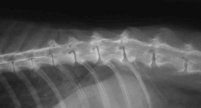
Diagnosis of a slipped disk is rarely possible using standard X-rays alone. A standard X-ray can only show the bones of the vertebrae and not the joints between them (the disks) or the spinal cord running inside them. Sometimes changes seen on conventional X-rays suggest disk degeneration without the animal showing any signs. A definite diagnosis of a slipped disk can only be made using either myelography (X-rays taken after the injection of dye around the spinal cord), CT (computed tomography) or MRI (magnetic resonance imaging). These special tests help to confirm if there is a slipped disk, where it is and will also show up other causes of spinal pain or paralysis if they are present.
Will my pet need an operation?
In most cases a slipped disk should be considered to be a surgical disease except where:
- This is the first time the animal has had back pain.
- The animal has a medical condition that contraindicates general anesthesia.
- Or if the animal has minimal spinal cord compression and it is suspected that spinal bruising is responsible for most of the signs.
Non-surgical treatment consists of strict rest, in a cage or room, depending on the size of your pet), for at least 4 weeks and treatment with drugs that will reduce inflammation and pain. Your veterinarian will want to see your pet regularly to ensure that they are not getting worse without surgery.
How can an operation help my pet?
Surgical treatment consists in drilling a hole in the vertebrae to remove the part of the IVD that is putting pressure on the spinal cord. Recovery time vary from 1 to 4 weeks. Despite carrying a small risk of causing further trauma, surgery should prevent further deterioration and relapse in the future. Success of surgery depends mainly on how much spinal cord function has been lost and especially whether or not and for how long the animal has lost the ability to feel pain in its toes. The prognosis is good for most animals that retain pain sensation. Paralyzed animals with no pain sensation in their rear legs have a slightly better than 50:50 chance of recovering the ability to walk unless this sensation has been lost for more than 48 hours when the prognosis become then very poor.
Will my pet recover without surgery?
Although surgical treatment is often preferred, cats with mild spinal cord compression or mostly bruising can recover with only rest and eventually anti-inflammatory medication. However those cats that do get to walk again, may take a long time to recover sometimes from 6 to 12 weeks.

